Optical ice thickness measurement in the VitroJet for time-efficient single particle structure determination
Rene J.M. Henderikx, Saba Shahzad, Maaike J.G. Schotman, Daniel Mann, Thomas V. Heidler, Dariush Ashtiani, Roger J.M. Jeurissen, Peter J. Peters, Carsten Sachse, Bart W.A.M.M. Beulen
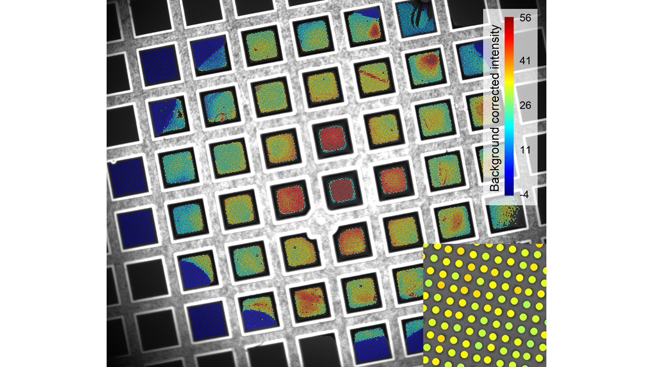
In cryo-electron microscopy structure determination, embedding biomolecules in vitreous ice of optimal thickness is critical. Ice thickness assessment and selection of suitable holes for data collection are currently part of time-consuming preparatory routines performed on expensive electron microscopes. For this reason, we developed a method to determine ice thickness during sample preparation using the optical camera integrated in the VitroJet. As a result, the ice thickness of buffer-suspended holes on an EM grid can be determined faithfully in the working range relevant for single particle cryo-EM. Dependent on the thickness, the error is below ± 20 nm in the range between 0 – 70 nm down to ± 10 nm in the thinnest ice regions (10 – 40 nm). Single particle structures of apoferritin were determined at two distinct thicknesses of 30 nm and 70 nm to demonstrate the suitability of optical layer thickness measurements for time-efficient cryo-EM structure determination.